

A new detector aims to reduce costs while improving the quality of nuclear medicine.
Physicians use nuclear medicine techniques such as SPECT scans to observe how the heart pumps, follow patterns of blood flow, and identify diseases that are otherwise hidden deep within the body. Current scanners, however, rely on detectors that are both costly and difficult to manufacture.
Researchers from Northwestern University and Soochow University in China have now developed the first perovskite-based detector capable of capturing individual gamma rays with exceptional precision for SPECT imaging. This innovation has the potential to make widely used nuclear imaging methods more accurate, efficient, affordable, and safe.
For patients, the benefits could include shorter scanning sessions, clearer diagnostic images, and lower exposure to radiation.
The study was recently published in the journal Nature Communications.
“Perovskites are a family of crystals best known for transforming the field of solar energy,” said Northwestern’s Mercouri Kanatzidis, the study’s senior author. “Now, they are poised to do the same for nuclear medicine. This is the first clear proof that perovskite detectors can produce the kind of sharp, reliable images that doctors need to provide the best care for their patients.”
“Our approach not only improves the performance of detectors but also could lower costs,” said co-corresponding author Yihui He, a professor at Soochow University. “That means more hospitals and clinics eventually could have access to the best imaging technologies.”
Kanatzidis is a Charles E. and Emma H. Morrison Professor of Chemistry at Northwestern’s Weinberg College of Arts and Sciences and a senior scientist at Argonne National Laboratory. Yihui He is a former postdoctoral fellow from Kanatzidis’ laboratory.
Why current detectors fall short
Nuclear medicine techniques such as SPECT (single-photon emission computing tomography) function like an invisible camera. A physician introduces a small, safe, and short-lived radiotracer into a targeted area of the patient’s body. This tracer releases gamma rays, which travel through tissues and are then captured by a detector outside the body. Each gamma ray acts like a pixel of light, and when millions of these pixels are recorded, computers assemble them into a three-dimensional image of organ activity.
Current detectors are typically made from cadmium zinc telluride (CZT) or sodium iodide (NaI), but both options come with drawbacks. CZT detectors are extremely costly, often ranging from hundreds of thousands to millions of dollars per camera, and the crystals are brittle, making them difficult to produce. NaI detectors are less expensive but bulkier, and they generate less precise images, similar to looking through a fogged window.
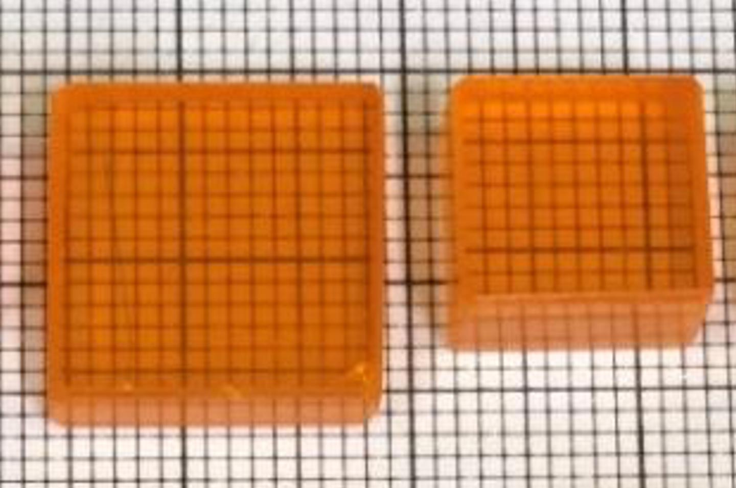
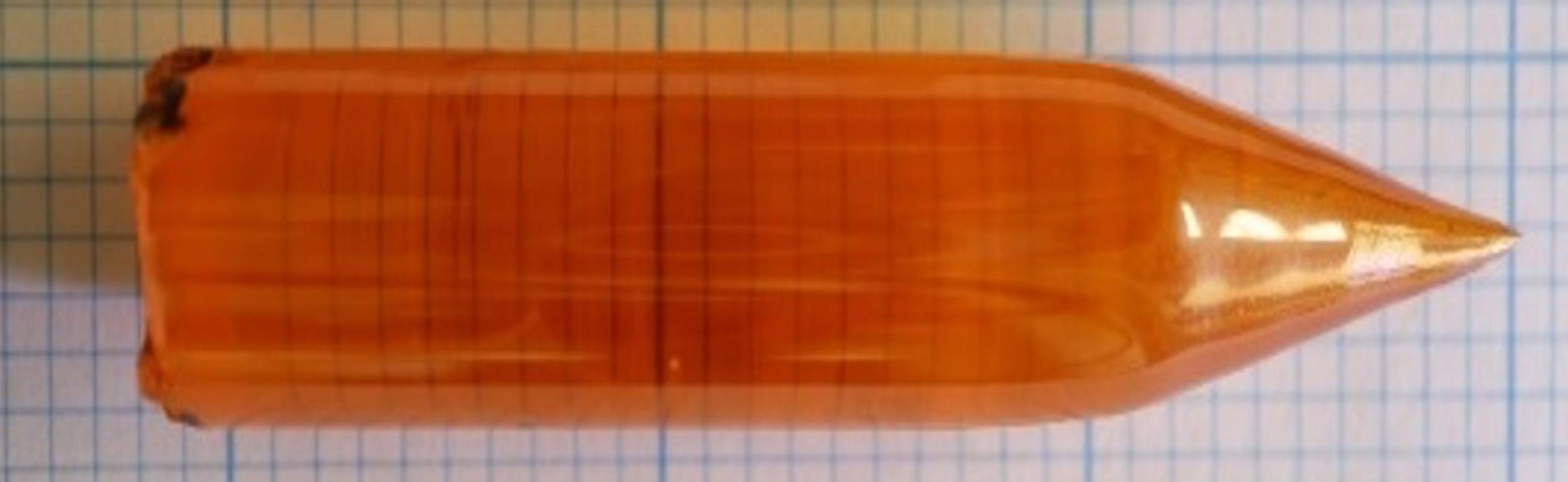
To address these challenges, the research team turned to perovskite crystals, which Kanatzidis has been studying for more than ten years. In 2012, his group created the first solid-film solar cells using perovskites. By 2013, he showed that single perovskite crystals could effectively detect X-rays and gamma rays. This advance, made possible through his team’s ability to grow high-quality crystals, set off a wave of international research and helped establish a new field focused on radiation detection materials.
Record-breaking imaging performance
“This work demonstrates how far we can push perovskite detectors beyond the laboratory,” Kanatzidis said. “When we first discovered in 2013 that perovskite single crystals could detect X-rays and gamma rays, we could only imagine their potential. Now, we’re showing that perovskite-based detectors can deliver the resolution and sensitivity needed for demanding applications like nuclear medicine imaging. It’s exciting to see this technology moving closer to real-world impact.”
Building on this foundation, Kanatzidis and He led the crystal growth, surface engineering and device design for the new study. By carefully growing and shaping these crystals, the researchers created a pixelated sensor — just like the pixels in a smartphone camera — that delivers record-breaking clarity and stability.
Leading the design and development of the prototype gamma-ray detector, He developed the camera’s pixelated architecture, optimized the multi-channel readout electronics, and carried out the high-resolution imaging experiments that validated the device’s capabilities. He, Kanatzidis, and their team demonstrated that perovskite-based detectors can achieve record energy resolutions and unprecedented single-photon imaging performance, paving the way for practical integration into next-generation nuclear medicine imaging systems.
Real-world impact and commercialization
“Designing this gamma-ray camera and demonstrating its performance has been incredibly rewarding,” He said. “By combining high-quality perovskite crystals with a carefully optimized pixelated detector and multi-channel readout system, we were able to achieve record-breaking energy resolution and imaging capabilities. This work shows the real potential of perovskite-based detectors to transform nuclear medicine imaging.”
In experiments, the detector was able to differentiate among gamma rays of different energies with the best resolution reported thus far. It also sensed extremely faint signals from a medical radiotracer (technetium-99m) commonly used in clinical practice and distinguished incredibly fine features, producing crisp images that could separate tiny radioactive sources spaced just a few millimeters apart. The detector also remained highly stable, collecting nearly all the tracer’s signal without loss or distortion. Because these new detectors are more sensitive, patients potentially could require shorter scan times or smaller doses of radiation.
Northwestern spinout company Actinia Inc. is commercializing this technology — working with partners in the medical device field to bring it out of the lab and into hospitals. Because they are easier to grow and use simpler components, perovskites offer a far less expensive alternative to CZT and NaI detectors without sacrificing quality. Perovskite-based detectors also offer a realistic pathway to imaging using a lower dose of a radiotracer than can be used with a NaI detector but at a price that ensures widespread patient access.
“Demonstrating that perovskites can deliver single-photon gamma-ray imaging is a milestone,” He said. “It shows these materials are ready to move beyond the laboratory and into technologies that directly benefit human health. From here, we see opportunities to refine the detectors further, scale up production, and explore entirely new directions in medical imaging.”
“High-quality nuclear medicine shouldn’t be limited to hospitals that can afford the most expensive equipment,” Kanatzidis said. “With perovskites, we can open the door to clearer, faster, safer scans for many more patients around the world. The ultimate goal is better scans, better diagnoses, and better care for patients.”
Reference: “Single photon γ-ray imaging with high energy and spatial resolution perovskite semiconductor for nuclear medicine” by Nannan Shen, Xuchang He, Tingting Gao, Bao Xiao, Yuquan Wang, Ruohan Ren, Haoming Qin, Khasim Saheb Bayikadi, Zhifu Liu, J. A. Peters, Bruce W. Wessels, Luyao Wang, Xiao Ouyang, Shuquan Wei, Qihao Sun, Xueping Liu, Yifei Lai, Xiaoping Ouyang, Zhifang Chai, Mercouri G. Kanatzidis and Yihui He, 30 August 2025, Nature Communications.
DOI: 10.1038/s41467-025-63400-7
Supported by the Defense Threat Reduction Agency (award number HDTRA12020002), the Consortium for Interaction of Ionizing Radiation with Matter University Research Alliance, the National Key R&D Program of China (award number 2021YFF0502600), the National Natural Science Foundation of China (award number U2267211) and the Jiangsu Natural Science Foundation (award number BK20240822).
Never miss a breakthrough: Join the SciTechDaily newsletter.